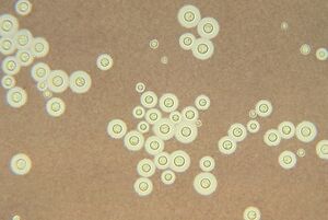
Radiotrophic fungus (nonfiction)
Radiotrophic fungi are fungi which perform radiosynthesis, using the pigment melanin to convert gamma radiation into chemical energy for growth.
Synopsis
The biological pathway for energy conversion may be similar to anabolic pathways for the synthesis of reduced organic carbon (e.g., carbohydrates) in phototrophic organisms, which convert photons from visible light with pigments such as chlorophyll whose energy is then used in photolysis of water to generate usable chemical energy (as ATP) in photophosphorylation or photosynthesis. However, whether melanin-containing fungi employ a similar multi-step pathway as photosynthesis, or some chemosynthesis pathways, is unknown [February 2020].
Observations
Discovery
Radiotrophic fungi were discovered in 1991 growing inside and around the Chernobyl Nuclear Power Plant.[1] Research at the Albert Einstein College of Medicine showed that three melanin-containing fungi—Cladosporium sphaerospermum, Wangiella dermatitidis, and Cryptococcus neoformans—increased in biomass and accumulated acetate faster in an environment in which the radiation level was 500 times higher than in the normal environment. Exposure of C. neoformans cells to these radiation levels rapidly (within 20–40 minutes of exposure) altered the chemical properties of its melanin, and increased melanin-mediated rates of electron transfer (measured as reduction of ferricyanide by NADH) three- to four-fold compared with unexposed cells.[2] Similar effects on melanin electron-transport capability were observed by the authors after exposure to non-ionizing radiation, suggesting that melanotic fungi might also be able to use light or heat radiation for growth.
Comparisons with non-melanized fungi
Melanization may come at some metabolic cost to the fungal cells. In the absence of radiation, some non-melanized fungi (that had been mutated in the melanin pathway) grew faster than their melanized counterparts. Limited uptake of nutrients due to the melanin molecules in the fungal cell wall or toxic intermediates formed in melanin biosynthesis have been suggested to contribute to this phenomenon. It is consistent with the observation that despite being capable of producing melanin, many fungi do not synthesize melanin constitutively (i.e., all the time), but often only in response to external stimuli or at different stages of their development. The exact biochemical processes in the suggested melanin-based synthesis of organic compounds or other metabolites for fungal growth, including the chemical intermediates (such as native electron donor and acceptor molecules) in the fungal cell and the location and chemical products of this process, are unknown.
"Ionizing Radiation Changes the Electronic Properties of Melanin and Enhances the Growth of Melanized Fungi"
"Ionizing Radiation Changes the Electronic Properties of Melanin and Enhances the Growth of Melanized Fungi" is a 2007 research paper by Ekaterina Dadachova, Ruth A. Bryan, Xianchun Huang, Tiffany Moadel, Andrew D. Schweitzer, Philip Aisen, Joshua D. Nosanchuk, and Arturo Casadevall.
Published online (2007)
Background
Melanin pigments are ubiquitous in nature. Melanized microorganisms are often the dominating species in certain extreme environments, such as soils contaminated with radionuclides, suggesting that the presence of melanin is beneficial in their life cycle. We hypothesized that ionizing radiation could change the electronic properties of melanin and might enhance the growth of melanized microorganisms.
Methodology/Principal Findings
Ionizing irradiation changed the electron spin resonance (ESR) signal of melanin, consistent with changes in electronic structure. Irradiated melanin manifested a 4-fold increase in its capacity to reduce NADH relative to non-irradiated melanin. HPLC analysis of melanin from fungi grown on different substrates revealed chemical complexity, dependence of melanin composition on the growth substrate and possible influence of melanin composition on its interaction with ionizing radiation. XTT/MTT assays showed increased metabolic activity of melanized C. neoformans cells relative to non-melanized cells, and exposure to ionizing radiation enhanced the electron-transfer properties of melanin in melanized cells. Melanized Wangiella dermatitidis and Cryptococcus neoformans cells exposed to ionizing radiation approximately 500 times higher than background grew significantly faster as indicated by higher CFUs, more dry weight biomass and 3-fold greater incorporation of 14C-acetate than non-irradiated melanized cells or irradiated albino mutants. In addition, radiation enhanced the growth of melanized Cladosporium sphaerospermum cells under limited nutrients conditions.
Conclusions/Significance
Exposure of melanin to ionizing radiation, and possibly other forms of electromagnetic radiation, changes its electronic properties. Melanized fungal cells manifested increased growth relative to non-melanized cells after exposure to ionizing radiation, raising intriguing questions about a potential role for melanin in energy capture and utilization.
References
- Science News, Dark Power: Pigment seems to put radiation to good use, Week of May 26, 2007; Vol. 171, No. 21 , p. 325 by Davide Castelvecchi
- Dadachova E, Bryan RA, Huang X, Moadel T, Schweitzer AD, Aisen P, Nosanchuk JD, Casadevall A (2007). Rutherford J (ed.). "Ionizing radiation changes the electronic properties of melanin and enhances the growth of melanized fungi". PLoS ONE. 2 (5): e457. Bibcode:2007PLoSO...2..457D. doi:10.1371/journal.pone.0000457. PMC 1866175. PMID 17520016.
- Calvo AM, Wilson RA, Bok JW, Keller NP (2002). "Relationship between secondary metabolism and fungal development". Microbiol Mol Biol Rev. 66 (3): 447–459. doi:10.1128/MMBR.66.3.447-459.2002. PMC 120793. PMID 12208999.
In the News
Fiction cross-reference
Nonfiction cross-reference
External links
- Radiotrophic fungus @ Wikipedia
- Ionizing Radiation Changes the Electronic Properties of Melanin and Enhances the Growth of Melanized Fungi @ National Institute of Health
- Einstein College of Medicine on radiotrophic fungi
- The blacker the better… especially in Chernobyl at Earthling Nature.

